Everything good had to happen and it did
By M S Nazki
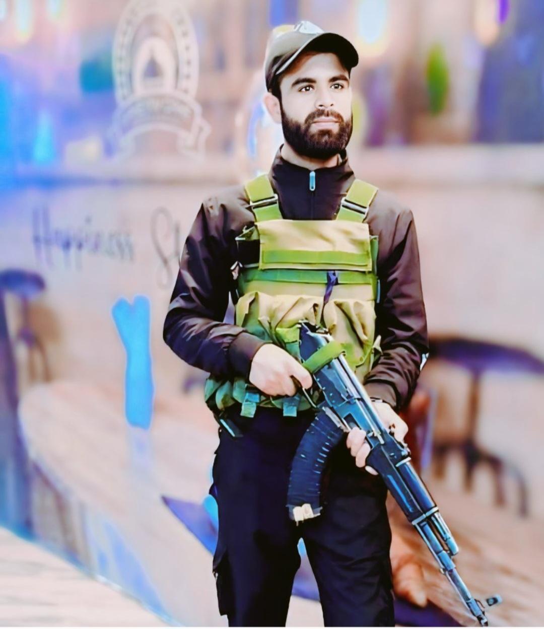
He did not have a face that could discern as to who he was and then entered the Fire & Fury Corps medical brigade! And what else was expected? Everything good had to happen and it did!
-For the soul, depression is an initiation, a rite of passage. If we think that depression, so empty and dull, is void of imagination, we may overlook its initiatory aspects. We may be imagining imagination itself from a point of view foreign to Saturn; emptiness can be rife with feeling-tone, images of catharsis, and emotions of regret and loss. As a shade of mood, gray can be as interesting and as variegated as it is in black-and-white photography. If”! The man who was undergoing the surgery must have found himself in such a predicament!
-But he carried hope along with him and the hope did arrive!
-In a remarkable feat of Maxillofacial Surgery at Fire and Fury Corps Dental Unit, a serving soldier with extensive PANFACIAL TRAUMA Secondary due to crush injury was successfully operated with Open Reduction and Internal Fixation in the extreme high altitude region of Leh.
-A remarkable feat wherein, patients with such grave injuries are generally transferred to higher centers. The patient was operated in situ uneventfully thus reducing morbidity, permanent facial disfigurement and saving precious life.
-Traumatic panfacial fracture repair is one of the most complex and challenging reconstructive procedures to perform. Several principles permeate throughout literature regarding the repair of panfacial injuries in a stepwise fashion. The primary goal of management in most of these approaches is to restore the occlusal relationship at the beginning of sequential repair so that other structures can fall into alignment. Through proper positioning of the occlusion and the mandibular-maxillary unit with the skull base, the spatial relationships and stability of midface buttresses and pillars can then be re-established. Here, the authors outline the sequencing of panfacial fracture repair for the restoration of anatomical relationships and the optimization of functional and structural outcomes.
-The approach for the treatment of panfacial injuries may seem difficult at first, but if a stepwise approach is followed with an understanding of the principles of repair, the outcome can be optimized. There are several mechanisms of injury which exist that propagate along the zones of weakness within the midface and mandible to represent a common fracture pattern. Standard fracture patterns are classically described by LeFort; yet generally, there is a combination of various components of the LeFort fractures and other fractures. The components of the true panfacial fracture include the lower third, the middle third, and upper third of the face, but the involvement of the midface and mandible constitute the same principles of repair, as a true panfacial fracture would dictate. The components of panfacial fractures are listed in Table 1. Usually, there is a combination of all of these various fractures.
-One of the primary concerns with regards to the repair of panfacial fractures is airway management. There are four established mechanisms for the airway: oral intubation, nasal intubation, submental intubation, and a tracheostomy.1 The latter three of these intubations allows for mandibular–maxillary fixation with full dentition. Oral intubation is possible when there is an absence of occlusion or absent teeth that allows the oral tube to be placed posteriorly in the mouth. Nasal intubation is often possible; however, with complex nasal and naso-orbito-ethmoid fractures, in addition to mandibular and palatal fractures, there is concern for postoperative management of the airway.2 There can be significant edema or packing within the nose in combination with mandibular–maxillary fixation that also leads to concern about maintaining airway patency. Submental intubation has been shown to be a safe approach with the tube out of the way, but the postoperative issues in regards to nasal packing and mandibular–maxillary fixation still exist. A tracheostomy allows the tube to be away from the structures being repaired and also has postoperative control of the airway. Of course, there are concerns about postoperative tracheostomy-related complications; however, the risk of tracheostomy is relatively low when compared with the risk of airway management postoperatively.
-Once the airway has been established, the repair of panfacial injuries follows a systematic approach. There are different philosophies about inside-out or bottom-up versus outside-in or top-down approaches. The inside-out thought process is reconstructing the maxillary–mandibular unit as the first major step and then focusing on the midface structures. This would allow the occlusal relationship to be restored and then “built out” from that process. The outside-in, or top-down, mentality would be reconstructing the outer facial frame and the bony pillars, such as the zygomatic arch and the frontal areas, and then addressing the interfacial frame.3 4 These two thought processes have permeated the literature and teaching for decades. In actuality, the best course of action is to follow a combined process. The primary goal would be to restore the occlusal relationship and then the spatial relationship between the occlusal structures and the skull base.
-Historically, the lines of weakness were first described by LeFort in 1901. This was followed by descriptions of the buttresses in 1916 by Cryer, and by illustrations of the vertical pillars and horizontal buttresses. Epsteen and Dingman described the palatine and maxillary fractures as important for structural stability of the midface; Ferre et al finalized the importance of this relationship to the cranial base. This led to our understanding of the anatomy and physiology of the bony structural components of the midface in relation to the skull base and the mandible.
Repair of these fractures was previously performed with wires and various methods of fixation techniques, which led to widened and flattened facies. With the advent of rigid fixation in the 1960s, there has been a significant evolution of the principles for restoring the structural relationships between the occlusal services and the skull base, in addition to narrowing the midface appropriately. Currently, there are further soft tissue repair refinements that have led to optimal outcomes.The consequences of suboptimal repair would involve malocclusion with a loss of the structural relationship between the mandible and the midface.
The components of panfacial fractures are outlined in Table 1. The definition of panfacial fracture incorporates the lower-third, middle-third, and upper-third facial components usually in a combination of fractures. There are multiple buttresses within the midface that need to be approached to restore the midface height, midface projection, and midface width, in addition to restoring the occlusal relationship. The medial buttresses are along the nasal frontal bone to the anterior maxillary alveolus. The lateral zygomatic maxillary buttresses extend along the zygoma and malar bone to the lateral maxillary alveolus . The pterygomaxillary buttress has a medial component that extends from the posterior alveolus and palate to the cranial base, and a lateral component that extends from the lateral pterygoid plate to the greater wing and lateral wall of the sphenoid. There is a central sphenovomerine buttress, which is along the central posterior palate to the floor of the sphenoid sinus
With disruption of all of the buttresses and the occlusal relationship, there is a tendency for facial widening, flattening, and rotation of the maxilla. Consequently, there will be an appearance of an obtuse nasolabial angle with impaction of the midface, which will seem like an open-bite deformity. The loss of bony relationships along the sphenoid bone or the lateral walls of the orbit with fractures along the zygomatic arch will result in apparent facial widening.With condylar fractures or ramus fractures, there is also a collapse of the mandibular relationship at the skull base.Therefore, the primary treatment goal is to approach these fractures in a stepwise fashion with proper sequencing of repairs by restoring the occlusal relationship and extending out to the repair of all of the buttresses.
Sequencing: The key to sequencing in panfacial fracture management is to understand both the principles of buttress reconstruction and the need for restoring the spatial relationship of the occlusion in the skull base.10 With panfacial fractures, there is a compromise of the mandibular–maxillary unit and the relationship between these two structures and the skull base. The midface is also violated with the loss of key components necessary for anatomical alignment. For example, the repair of mandibular–maxillary fractures often will rely on the stable structure of the upper face and vice-versa. With panfacial fractures, there is a loss of the customary structures for anatomical alignment.
Palate fractures need to be closed and held in position. This example depicts opening the fracture and placing a three-dimensional plate. The fracture may rotate slightly, but will not distract. Mandibular–maxillary fixation will restore the occlusion.
The patient’s head is held in position and the Rowe disimpaction forceps are placed in the mouth and nose. The maxilla is pulled out. Care must be taken to avoid injury to the skull base and propagation of a cerebrospinal leak. After the palate is repaired, the mandible will need to be corrected with arch bars. There is the standard parasymphyseal and angle-type fracture of the mandible, but in patients with panfacial fractures, there may be more comminuted segments of the condyle on either or both sides.With significant fractures to the condyles, loss of the spatial relationship between the mandible and skull base will occur. To address this abnormality, the mandible would be restored to its premorbid condition through mandibular–maxillary fixation and restoration of the occlusion. The goal would be to have the LeFort I level component of the maxilla set with the mandible as a single “block” that would then articulate with the skull base, restoring the spatial relationships between these structures.
An external approach to mandibular repair should be strongly considered in patients with panfacial trauma as they will often have multiple other injuries and require care in the intensive care unit. Oral care after mandibular repair may be somewhat tenuous postoperatively, especially in patients with other morbidities, with higher chances of intraoral dehiscence. Therefore, the severity of injuries is not an absolute indication to perform an external repair, but consideration should be given
Mandible fractures are normally repaired via intraoral incisions. With severe facial trauma, external incisions facilitate the repair and also minimize the risk of intraoral dehiscence. The lingual cortex is readily seen and reduced.
Intracondylar fractures can be treated in a normal fashion with closed treatment for functional restoration. Ordinarily, a condyle fracture can be treated closed if there is a stable structural relationship with the midface; however, in panfacial fractures, this is not present. Hence, lower fractures such as subcondylar or low condylar neck fractures often call for open treatment. These can be approached by either endoscopic or retromandibular repair.14 Once the mandible and the maxilla have been repaired, in addition to restoration of the vertical height, this entire unit will be equivalent to a block that will then be able to articulate and provide stable foundation for the repair of the midface. At this point, the repair of the panfacial fracture can be staged . There is no commitment for the repair of the midface because it may take several hours to repair the palate and to restore the mandible and vertical height.
The mandible and maxilla need to be restored prior to the mid- and upper-face correction. This example shows the mandibular–maxillary unit working together en bloc.
The subcondylar fracture can be repaired to restore the spatial relationship with the skull base. The image shows repair of the subcondylar fracture via a retromandibular approach.
The middle-third and upper-third facial fracture repairs are done via anterior and posterior approaches. The posterior approach (using a coronal incision) would either be curvilinear or a stealth-type incision.15 The advantages of the stealth incision would be that it would blend within the hairline and stagger the layered hair. It also facilitates closure, as the interstices of the incision are easy to close. The curvilinear incision is more expedient and also lends itself to secondary correction in an easier fashion. Patients that have anterior baldness can have the incision performed posteriorly to the occipital region, so that the scar will be hidden by hair. The standard anterior approach would be for transpalpebral incisions. These would either be transcutaneous or transconjunctival. If a transconjunctival incision is made, a lateral canthotomy will need to be made to have full exposure to the inferior rim. Upper buccal sulcus incisions will also need to be made for the repair of the LeFort I.
Through the coronal incision, the zygomatic arches will need to be exposed if repair is required.10 Care will need to be taken to dissect along the deep temporal fascia and to elevate the superficial temporal fascia to avoid injury to the frontal branch of the facial nerve. The frontal branch may have some weakness postoperatively, but this is usually a traction injury and will likely recover over time. The dissection over the deep temporal fascia is performed in a posterior to anterior fashion to dissect along the galeotemporal plane and release this galeotemporal fusion point to avoid injury to the frontal branch of the facial nerve.The frontal bar and the lateral rims can then be effectively exposed. The zygomatic arch will be exposed by creating a leaflet of tissue, ∼ 1.5 cm superior to the arch. This will facilitate closure at the completion of the case when the fascia will need to be resuspended.
With the naso-orbito-ethmoid components of the fracture, the dissection will need to be performed through the medial portion of the nasal frontal region.Care must be taken to dissect along the nasal frontal region down into the superior orbital area at the superior edge of the ethmoid. The superior trochlea is in this region. If there are small bone fragments in this region, the trochlea may be attached to these fragments; it is safer to just allow these fragments to release and not to disturb them because damage to the superior trochlea may lead to superior oblique muscle dysfunction. Once the tissue is closed upon completion of the procedure, those fragments will lay back down to position and scar down to maintain the function of the superior trochlea. The medial dissection is then performed along the ethmoid, if there is a nasoethmoid fracture component. All of the fractures will need to be exposed before there can be any reduction and subsequent fixation.
At this point, the occlusion and mandibular–maxillary fixation has been reestablished with intact spatial relationships to the skull base. This will assist in restoring the vertical facial height. The malar bones through the LeFort III level will need to be aligned. The zygomaticosphenoid junction, which is the lateral wall of the orbit, is a wide bone component and usually is not comminuted. This structure is the key to the anatomical alignment of the zygomaticomaxillary complex and can be exposed by two major methods Tissue can be reflected from the temporalis muscle to expose the zygomaticosphenoid junction from a posterolateral approach or the orbit can be entered and the globe can be reflected medially with the junction seen at that point. This is a wide bone contact point that allows for restoration of the anterolateral positioning of the zygomatic complex
The frontozygomatic bone has the strongest point of fixation, but is least reliable for reduction. The lateral wall (zygomaticosphenoid junction) is plated. This is the key to anatomical reduction of a complicated zygomaticomaxillary complex fracture and can be held in reduction with a plate, as shown.
The other area that is difficult to repair is along the zygomatic process, or the zygomatic arch, of the temporal bone if it is sheared off. The zygomatic process of the temporal bone can be visualized by releasing the temporalis muscle posteriorly. This segment is pushed back and held into reduction. A 10-mm-length screw can be passed through this and provide stable fixation and restoration of the posterior portion of the zygomatic arch . With restoration of both the zygomaticosphenoid and the zygomatic process of the temporal bone, the bony fragments will begin to align. The zygomatic arch can be held together with a plate because it is somewhat flat in the central region with a gentle bend anteriorly and posteriorly. Quite often, when the inferior rim is viewed after restoration of the zygomaticosphenoid and the zygomaticotemporal components, there is stable positioning of the lateral portion of the inferior rim. With the midface reduced, the medial component of the infraorbital rim is rigidly fixed.
Once the midface is largely restored, the areas that will need to be addressed in the final stages of repair include the naso-orbito-ethmoid component, the orbital floor, and the LeFort I-level repairs. The naso-orbito-ethmoid region will often be fractured in three types. The type I fracture can be simply plated, but the type II fracture will need wiring. Type III fractures will need bone grafting and wiring.2 Bone graft is readily harvested from the parietal skull and is used liberally for the reconstruction of the nasal dorsum, the medial orbital wall, and the naso-orbito-ethmoid region. Once the naso-orbito-ethmoid area has been repaired, the medial canthal tendons are reattached and tightened down . The LeFort I level repair has the highest degree of forgiveness with regards to anatomical alignment. As noted, there are four buttresses in the area where the LeFort I level is plated. There are the lateral and medial zygomatic and maxillary buttresses for both the right and left sides. Care will need to be taken to avoid screws through the two-thirds, but sometimes this is unavoidable in significant fractures. If there is significant comminution in this area, bone grafting is utilized.4 Lastly, the orbital floors are typically the final areas to be repaired.Usually, porous polyethylene is of little use in the area because it tends to be soft and may bow slightly in the orbital floor. With panfacial fractures, the orbital floor is generally comminuted and a more stable construct will need to be used, either with a titanium prosthetic or bone grafting. There are commercially available hybrid implants with titanium and porous polyethylene that may also be considered.
Closure and Recovery:
At this point, the face should be completely reconstructed. The mandibular–maxillary fixation will need to be released to assess the occlusion and make sure there is central occlusion with maximum intercuspation . The forced duction test will also need to be performed to verify freely moving orbital cone contents.This will all need to be done in anticipation of closure. The closure of the midface is a very important aspect of panfacial trauma management, so even part of the opening has to do with the anticipation of the closure. The intraoral incisions are closed in the usual fashion with reapproximating the muscle and then the mucosa. This could either be with running sutu